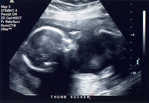
Any sound with a frequency above 20 000 Hz (or 20 kHz)—that is, above the highest audible frequency—is defined to be ultrasound. In practice, it is possible to create ultrasound frequencies up to more than a gigahertz. (Higher frequencies are difficult to create; furthermore, they propagate poorly because they are very strongly absorbed.) Ultrasound has a tremendous number of applications, which range from burglar alarms to use in cleaning delicate objects to the guidance systems of bats. We begin our discussion of ultrasound with some of its applications in medicine, in which it is used extensively both for diagnosis and for therapy.
The characteristics of ultrasound, such as frequency and intensity, are wave properties common to all types of waves. Ultrasound also has a wavelength that limits the fineness of detail it can detect. This characteristic is true of all waves. We can never observe details significantly smaller than the wavelength of our probe; for example, we will never see individual atoms with visible light, because the atoms are so small compared with the wavelength of light.
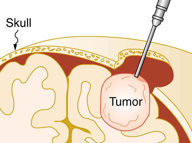
Ultrasound, like any wave, carries energy that can be absorbed by the medium carrying it, producing effects that vary with intensity. When focused to intensities of $10^{3}$ to $10^{5} {\text{W/m}}^{2}$ , ultrasound can be used to shatter gallstones or pulverize cancerous tissue in surgical procedures. (See [Figure 2].) Intensities this great can damage individual cells, variously causing their protoplasm to stream inside them, altering their permeability, or rupturing their walls through cavitation. Cavitation is the creation of vapor cavities in a fluid—the longitudinal vibrations in ultrasound alternatively compress and expand the medium, and at sufficient amplitudes the expansion separates molecules. Most cavitation damage is done when the cavities collapse, producing even greater shock pressures.
Most of the energy carried by high-intensity ultrasound in tissue is converted to thermal energy. In fact, intensities of $10^{3}$ to $10^{4} {\text{W/m}}^{2}$ are commonly used for deep-heat treatments called ultrasound diathermy. Frequencies of 0.8 to 1 MHz are typical. In both athletics and physical therapy, ultrasound diathermy is most often applied to injured or overworked muscles to relieve pain and improve flexibility. Skill is needed by the therapist to avoid “bone burns” and other tissue damage caused by overheating and cavitation, sometimes made worse by reflection and focusing of the ultrasound by joint and bone tissue.
In some instances, you may encounter a different decibel scale, called the sound pressure level, when ultrasound travels in water or in human and other biological tissues. We shall not use the scale here, but it is notable that numbers for sound pressure levels range 60 to 70 dB higher than you would quote for $\beta$ , the sound intensity level used in this text. Should you encounter a sound pressure level of 220 decibels, then, it is not an astronomically high intensity, but equivalent to about 155 dB—high enough to destroy tissue, but not as unreasonably high as it might seem at first.
When used for imaging, ultrasonic waves are emitted from a transducer, a crystal exhibiting the piezoelectric effect (the expansion and contraction of a substance when a voltage is applied across it, causing a vibration of the crystal). These high-frequency vibrations are transmitted into any tissue in contact with the transducer. Similarly, if a pressure is applied to the crystal (in the form of a wave reflected off tissue layers), a voltage is produced which can be recorded. The crystal therefore acts as both a transmitter and a receiver of sound. Ultrasound is also partially absorbed by tissue on its path, both on its journey away from the transducer and on its return journey. From the time between when the original signal is sent and when the reflections from various boundaries between media are received, (as well as a measure of the intensity loss of the signal), the nature and position of each boundary between tissues and organs may be deduced.
Reflections at boundaries between two different media occur because of differences in a characteristic known as the acoustic impedance $Z$ of each substance. Impedance is defined as
where $\rho$ is the density of the medium (in $\text{kg/m}^{3}$ ) and $v$ is the speed of sound through the medium (in m/s). The units for $Z$ are therefore $\text{kg/m}^{2}·\text{s}$.
[Table 1] shows the density and speed of sound through various media (including various soft tissues) and the associated acoustic impedances. Note that the acoustic impedances for soft tissue do not vary much but that there is a big difference between the acoustic impedance of soft tissue and air and also between soft tissue and bone.
| Medium | Density (kg/m3) | Speed of Ultrasound (m/s) | Acoustic Impedance $$\left(\text{kg/}\left({\text{m}}^{2}\cdot \text{s}\right)\right) $$ |
|---|---|---|---|
| Air | 1.3 | 330 | $$429 $$ |
| Water | 1000 | 1500 | $$1.5 \times 10^{6} $$ |
| Blood | 1060 | 1570 | $$1.66 \times 10^{6} $$ |
| Fat | 925 | 1450 | $$1.34 \times 10^{6} $$ |
| Muscle (average) | 1075 | 1590 | $$1.70 \times 10^{6} $$ |
| Bone (varies) | 1400–1900 | 4080 | $$5.7 \times 10^{6} $$ to $$7.8 \times 10^{6} $$ |
| Barium titanate (transducer material) | 5600 | 5500 | $$30.8 \times 10^{6} $$ |
At the boundary between media of different acoustic impedances, some of the wave energy is reflected and some is transmitted. The greater the difference in acoustic impedance between the two media, the greater the reflection and the smaller the transmission.
The intensity reflection coefficient $a$ is defined as the ratio of the intensity of the reflected wave relative to the incident ( transmitted) wave. This statement can be written mathematically as
where ${Z}_{1}$ and ${Z}_{2}$ are the acoustic impedances of the two media making up the boundary. A reflection coefficient of zero (corresponding to total transmission and no reflection) occurs when the acoustic impedances of the two media are the same. An impedance “match” (no reflection) provides an efficient coupling of sound energy from one medium to another. The image formed in an ultrasound is made by tracking reflections (as shown in [Figure 3]) and mapping the intensity of the reflected sound waves in a two-dimensional plane.
(a) Using the values for density and the speed of ultrasound given in [Table 1], show that the acoustic impedance of fat tissue is indeed $1.34 \times 10^{6} \text{kg/m}^{2}·\text{s}$.
(b) Calculate the intensity reflection coefficient of ultrasound when going from fat to muscle tissue.
Strategy for (a)
The acoustic impedance can be calculated using $Z=\text{ρv}$ and the values for $\rho$ and $v$ found in [Table 1].
Solution for (a)
(1) Substitute known values from [Table 1] into $Z= \rho v$.
(2) Calculate to find the acoustic impedance of fat tissue.
This value is the same as the value given for the acoustic impedance of fat tissue.
Strategy for (b)
The intensity reflection coefficient for any boundary between two media is given by $a=\frac{ {\left({Z}_{2}-{Z}_{1}\right)}^{2}}{ {\left({Z}_{1} + {Z}_{2}\right)}^{2}}$ , and the acoustic impedance of muscle is given in [Table 1].
Solution for (b)
Substitute known values into $a=\frac{ {\left({Z}_{2}-{Z}_{1}\right)}^{2}}{ {\left({Z}_{1} + {Z}_{2}\right)}^{2}}$ to find the intensity reflection coefficient:
Discussion
This result means that only 1.4% of the incident intensity is reflected, with the remaining being transmitted.
The applications of ultrasound in medical diagnostics have produced untold benefits with no known risks. Diagnostic intensities are too low (about $10^{-2} {\text{W/m}}^{2}$ ) to cause thermal damage. More significantly, ultrasound has been in use for several decades and detailed follow-up studies do not show evidence of ill effects, quite unlike the case for X-rays.
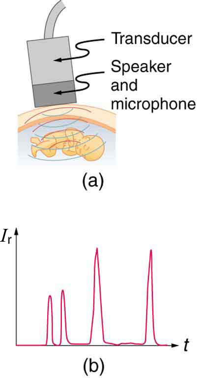
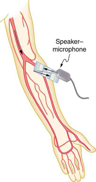
The most common ultrasound applications produce an image like that shown in [Figure 4]. The speaker-microphone broadcasts a directional beam, sweeping the beam across the area of interest. This is accomplished by having multiple ultrasound sources in the probe’s head, which are phased to interfere constructively in a given, adjustable direction. Echoes are measured as a function of position as well as depth. A computer constructs an image that reveals the shape and density of internal structures.
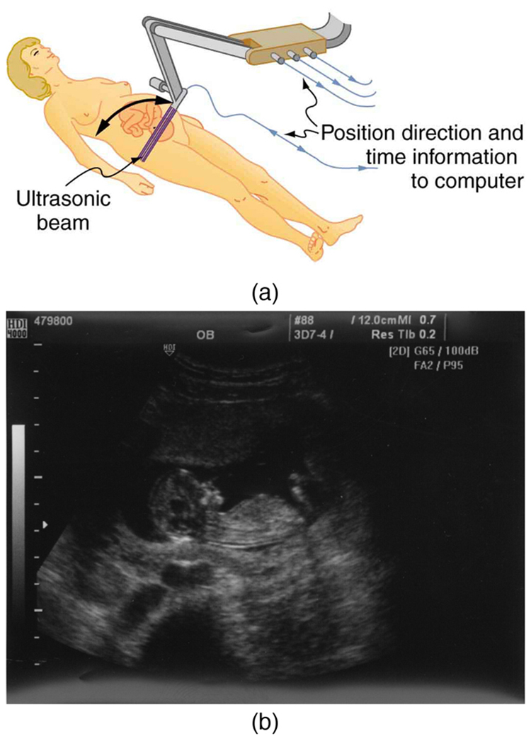
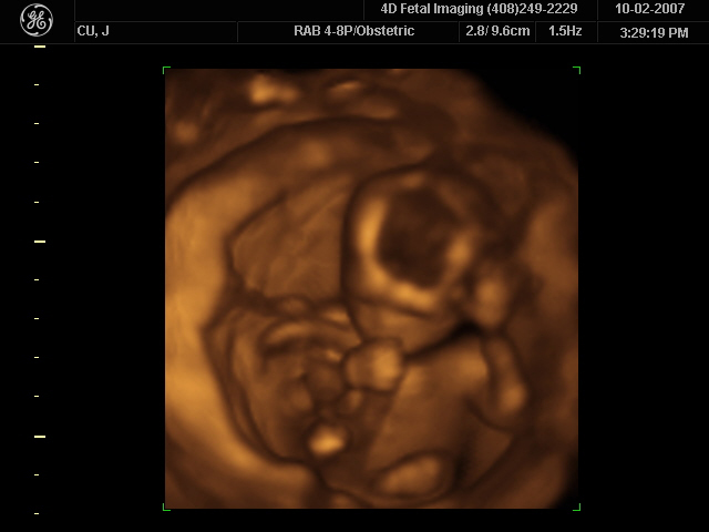
How much detail can ultrasound reveal? The image in [Figure 4] is typical of low-cost systems, but that in [Figure 5] shows the remarkable detail possible with more advanced systems, including 3D imaging. Ultrasound today is commonly used in prenatal care. Such imaging can be used to see if the fetus is developing at a normal rate, and help in the determination of serious problems early in the pregnancy. Ultrasound is also in wide use to image the chambers of the heart and the flow of blood within the beating heart, using the Doppler effect (echocardiology).
Whenever a wave is used as a probe, it is very difficult to detect details smaller than its wavelength $\lambda$ . Indeed, current technology cannot do quite this well. Abdominal scans may use a 7-MHz frequency, and the speed of sound in tissue is about 1540 m/s—so the wavelength limit to detail would be
$\lambda =\frac{ {v}_{w}}{f}=\frac{1540 \text{m/s}}{7 \times 10^{6} \text{Hz}}=0.22 \text{mm}$ . In practice, 1-mm detail is attainable, which is sufficient for many purposes. Higher-frequency ultrasound would allow greater detail, but it does not penetrate as well as lower frequencies do. The accepted rule of thumb is that you can effectively scan to a depth of about $500\lambda$ into tissue. For 7 MHz, this penetration limit is $500×0.22 \text{mm}$ , which is 0.11 m. Higher frequencies may be employed in smaller organs, such as the eye, but are not practical for looking deep into the body.
In addition to shape information, ultrasonic scans can produce density information superior to that found in X-rays, because the intensity of a reflected sound is related to changes in density. Sound is most strongly reflected at places where density changes are greatest.
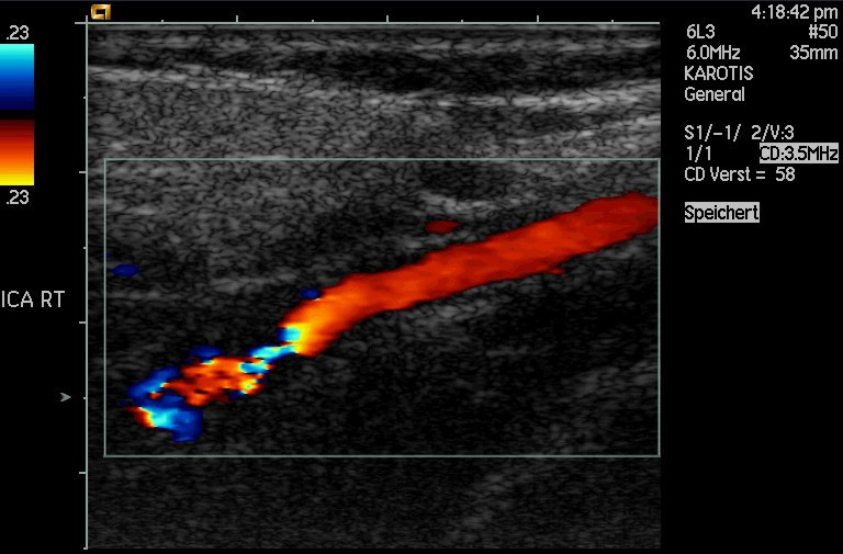
Another major use of ultrasound in medical diagnostics is to detect motion and determine velocity through the Doppler shift of an echo, known as Doppler-shifted ultrasound. This technique is used to monitor fetal heartbeat, measure blood velocity, and detect occlusions in blood vessels, for example. ( See [Figure 6].) The magnitude of the Doppler shift in an echo is directly proportional to the velocity of whatever reflects the sound. Because an echo is involved, there is actually a double shift. The first occurs because the reflector (say a fetal heart) is a moving observer and receives a Doppler-shifted frequency. The reflector then acts as a moving source, producing a second Doppler shift.
A clever technique is used to measure the Doppler shift in an echo. The frequency of the echoed sound is superimposed on the broadcast frequency, producing beats. The beat frequency is ${F}_{B}=\mid {f}_{1}-{f}_{2}\mid$ , and so it is directly proportional to the Doppler shift ( ${f}_{1}-{f}_{2}$ ) and hence, the reflector’s velocity. The advantage in this technique is that the Doppler shift is small (because the reflector’s velocity is small), so that great accuracy would be needed to measure the shift directly. But measuring the beat frequency is easy, and it is not affected if the broadcast frequency varies somewhat. Furthermore, the beat frequency is in the audible range and can be amplified for audio feedback to the medical observer.
Doppler-shifted radar echoes are used to measure wind velocities in storms as well as aircraft and automobile speeds. The principle is the same as for Doppler-shifted ultrasound. There is evidence that bats and dolphins may also sense the velocity of an object (such as prey) reflecting their ultrasound signals by observing its Doppler shift.
Ultrasound that has a frequency of 2.50 MHz is sent toward blood in an artery that is moving toward the source at 20.0 cm/s, as illustrated in [Figure 7. Use the speed of sound in human tissue as 1540 m/s. (Assume that the frequency of 2.50 MHz is accurate to seven significant figures.)
Strategy
The first two questions can be answered using ${f}_{\text{obs}}={f}_{s}\left( \frac{ {v}_{w}}{ {v}_{w} ± {v}_{s}}\right)$ and ${f}_{\text{obs}}={f}_{s}\left(\frac{ {v}_{w} ± {v}_{\text{obs}}}{ {v}_{w}}\right)$ for the Doppler shift. The last question asks for beat frequency, which is the difference between the original and returning frequencies.
Solution for (a)
(1) Identify knowns:
(2) Enter the given values into the equation.
(3) Calculate to find the frequency: 2 500 325 Hz.
Solution for (b)
(1) Identify knowns:
${f}_{obs}$ is the frequency received by the speaker-microphone.
The source velocity is ${v}_{b}$ .
The minus sign is used because the motion is toward the observer.
(2) Enter the given values into the equation:
(3) Calculate to find the frequency returning to the source: 2 500 649 Hz.
Solution for (c)
(1) Identify knowns:
(2) Substitute known values:
(3) Calculate to find the beat frequency: 649 Hz.
Discussion
The Doppler shifts are quite small compared with the original frequency of 2.50 MHz. It is far easier to measure the beat frequency than it is to measure the echo frequency with an accuracy great enough to see shifts of a few hundred hertz out of a couple of megahertz. Furthermore, variations in the source frequency do not greatly affect the beat frequency, because both ${f}_{s}$ and ${f}_{\text{obs}}$ would increase or decrease. Those changes subtract out in ${f}_{B}=\mid {f}_{\text{obs}}-{f}_{s}\mid .$
Industrial, retail, and research applications of ultrasound are common. A few are discussed here. Ultrasonic cleaners have many uses. Jewelry, machined parts, and other objects that have odd shapes and crevices are immersed in a cleaning fluid that is agitated with ultrasound typically about 40 kHz in frequency. The intensity is great enough to cause cavitation, which is responsible for most of the cleansing action. Because cavitation-produced shock pressures are large and well transmitted in a fluid, they reach into small crevices where even a low-surface-tension cleaning fluid might not penetrate.
Sonar is a familiar application of ultrasound. Sonar typically employs ultrasonic frequencies in the range from 30.0 to 100 kHz. Bats, dolphins, submarines, and even some birds use ultrasonic sonar. Echoes are analyzed to give distance and size information both for guidance and finding prey. In most sonar applications, the sound reflects quite well because the objects of interest have significantly different density than the medium in which they travel. When the Doppler shift is observed, velocity information can also be obtained. Submarine sonar can be used to obtain such information, and there is evidence that some bats also sense velocity from their echoes.
Similarly, there are a range of relatively inexpensive devices that measure distance by timing ultrasonic echoes. Many cameras, for example, use such information to focus automatically. Some doors open when their ultrasonic ranging devices detect a nearby object, and certain home security lights turn on when their ultrasonic rangers observe motion. Ultrasonic “measuring tapes” also exist to measure such things as room dimensions. Sinks in public restrooms are sometimes automated with ultrasound devices to turn faucets on and off when people wash their hands. These devices reduce the spread of germs and can conserve water.
Ultrasound is used for nondestructive testing in industry and by the military. Because ultrasound reflects well from any large change in density, it can reveal cracks and voids in solids, such as aircraft wings, that are too small to be seen with X-rays. For similar reasons, ultrasound is also good for measuring the thickness of coatings, particularly where there are several layers involved.
Basic research in solid state physics employs ultrasound. Its attenuation is related to a number of physical characteristics, making it a useful probe. Among these characteristics are structural changes such as those found in liquid crystals, the transition of a material to a superconducting phase, as well as density and other properties.
These examples of the uses of ultrasound are meant to whet the appetites of the curious, as well as to illustrate the underlying physics of ultrasound. There are many more applications, as you can easily discover for yourself.
Why is it possible to use ultrasound both to observe a fetus in the womb and also to destroy cancerous tumors in the body?
Ultrasound can be used medically at different intensities. Lower intensities do not cause damage and are used for medical imaging. Higher intensities can pulverize and destroy targeted substances in the body, such as tumors.
$\rho$ is the density of a medium through which the sound travels and $v$ is the speed of sound through that medium.
If audible sound follows a rule of thumb similar to that for ultrasound, in terms of its absorption, would you expect the high or low frequencies from your neighbor’s stereo to penetrate into your house? How does this expectation compare with your experience?
Higher frequencies are more readily absorbed than lower frequencies. Therefore, you would expect the low frequencies (the bass) from a neighbor’s stereo to penetrate into your house more effectively than the high frequencies.
This expectation matches common experience. It is often the case that you can hear the “thump-thump” of the bass from a neighbor’s stereo, while the higher-frequency melody and vocals are muffled or inaudible.
Elephants and whales are known to use infrasound to communicate over very large distances. What are the advantages of infrasound for long distance communication?
The primary advantage of infrasound for long-distance communication is its low rate of absorption. Lower-frequency sounds are less readily absorbed by the medium they travel through (like water or air) than higher-frequency sounds. This allows infrasound waves to travel much longer distances before their energy dissipates, making them ideal for communication over many kilometers.
It is more difficult to obtain a high-resolution ultrasound image in the abdominal region of someone who is overweight than for someone who has a slight build. Explain why this statement is accurate.
This statement is accurate because ultrasound waves are attenuated (absorbed and scattered) as they pass through tissue. In an overweight person, the ultrasound waves must travel through a thicker layer of subcutaneous fat to reach the abdominal organs. Fat tissue absorbs ultrasound energy, so the signal is weakened on its way to the organs and again on its way back to the transducer. This results in a weaker echo and a lower signal-to-noise ratio, which degrades the quality and resolution of the image.
Suppose you read that 210-dB ultrasound is being used to pulverize cancerous tumors. You calculate the intensity in watts per centimeter squared and find it is unreasonably high ( $10^{5} {\text{W/cm}}^{2}$ ). What is a possible explanation?
A possible explanation is that the 210-dB value is being reported using the sound pressure level (SPL) scale, which is commonly used in medical and underwater acoustics, rather than the sound intensity level (SIL) scale used in this textbook. The SPL scale uses a different reference pressure and can result in decibel values that are 60-70 dB higher than the SIL for the same sound wave in tissue.
Therefore, a 210-dB SPL is equivalent to roughly $210 - 65 = 145$ dB on the SIL scale. An intensity level of 145 dB is still very high, but it is a much more reasonable value for therapeutic ultrasound used to destroy tissue.
Unless otherwise indicated, for problems in this section, assume that the speed of sound through human tissues is 1540 m/s.
What is the sound intensity level in decibels of ultrasound of intensity $10^{5} {\text{W/m}}^{2}$ , used to pulverize tissue during surgery?
Strategy
The sound intensity level in decibels is calculated using the logarithmic formula $\beta \left(\text{dB}\right)=10{\text{log}}_{10}\left(\frac{I}{{I}_{0}}\right)$ , where ${I}_{0}=10^{-12} {\text{W/m}}^{2}$ is the threshold of hearing. This relationship allows us to express the enormous range of sound intensities encountered in medical applications on a manageable scale.
Solution
Identify the known values:
Substitute into the decibel formula:
Using the logarithm property ${\text{log}}_{10}\left(10^{x}\right)=x$ :
Discussion
This intensity level of 170 dB represents an extremely high energy concentration, approximately $10^{17}$ times greater than the threshold of human hearing. Such intensities are used therapeutically to destroy unwanted tissue through several mechanisms: direct mechanical disruption of cellular structures, cavitation (formation and violent collapse of vapor bubbles), and localized heating. This intensity is about $10^{10}$ times greater than diagnostic ultrasound (typically $10^{-2} {\text{W/m}}^{2}$ or 103 dB), ensuring that imaging procedures are safe while therapeutic applications can achieve the desired tissue destruction. The focused nature of therapeutic ultrasound allows surgeons to target specific tissues (such as gallstones or tumors) while minimizing damage to surrounding healthy tissue. At these intensities, exposure time must be carefully controlled to prevent unintended damage.
Is 155-dB ultrasound in the range of intensities used for deep heating? Calculate the intensity of this ultrasound and compare this intensity with values quoted in the text.
Strategy
We need to convert the decibel level to intensity using the inverse of the decibel formula. The relationship is $I={I}_{0}\times 10^{\beta /10}$ , where $\beta$ is in decibels and ${I}_{0}=10^{-12} {\text{W/m}}^{2}$ . We can then compare this calculated intensity to the range stated in the text for ultrasound diathermy (deep-heat treatments).
Solution
Start with the decibel formula and solve for intensity:
Divide both sides by 10:
Take the antilogarithm (raise 10 to both sides):
Solve for $I$ :
Substitute $\beta =155 \text{ dB}$ and ${I}_{0}=10^{-12} {\text{W/m}}^{2}$ :
Discussion
Yes, 155-dB ultrasound is indeed in the appropriate range for deep-heat treatments. The text states that ultrasound diathermy uses intensities of $10^{3}$ to $10^{4} {\text{W/m}}^{2}$ , and our calculated value of $3.16\times 10^{3} {\text{W/m}}^{2}$ falls right in the middle of this range. This intensity is high enough to deposit significant thermal energy deep within tissue (hence “deep heating”), causing the tissue temperature to rise by a few degrees Celsius. This heating effect increases blood flow, reduces muscle tension, and promotes healing—which is why ultrasound diathermy is commonly used in physical therapy and sports medicine for treating muscle injuries. The intensity is much lower than that used for tissue destruction ($10^{5} {\text{W/m}}^{2}$ ) but much higher than diagnostic imaging ($10^{-2} {\text{W/m}}^{2}$ ), placing it firmly in the therapeutic heating range. At these intensities, careful application by trained therapists is necessary to avoid “bone burns” and cavitation damage, particularly near joints where reflection and focusing can occur.
Find the sound intensity level in decibels of $2.00 \times 10^{-2} {\text{W/m}}^{2}$ ultrasound used in medical diagnostics.
Strategy
We use the decibel formula $\beta \left(\text{dB}\right)=10{\text{log}}_{10}\left(\frac{I}{{I}_{0}}\right)$ to convert the given intensity to a sound intensity level. The reference intensity is ${I}_{0}=10^{-12} {\text{W/m}}^{2}$ , the threshold of hearing at 1000 Hz.
Solution
Identify the known values:
Substitute into the decibel formula:
Use the logarithm property ${\text{log}}_{10}\left(a\times 10^{b}\right)={\text{log}}_{10}(a)+b$ :
Discussion
This intensity level of 103 dB represents the typical range for diagnostic medical ultrasound imaging, such as prenatal scans, echocardiography, and abdominal imaging. The intensity is $2.00\times 10^{10}$ times greater than the threshold of hearing, which might seem high, but it’s important to remember that ultrasound frequencies (typically 1-20 MHz) are far above the audible range and interact with tissue differently than audible sound. This diagnostic intensity is safe for routine medical use—it’s approximately $10^{7}$ times weaker than therapeutic ultrasound used for tissue destruction and about $100$ times weaker than ultrasound diathermy. Decades of clinical use with detailed follow-up studies have shown no harmful effects at these diagnostic intensities. The energy is too low to cause significant heating or cavitation damage, making ultrasound imaging one of the safest medical imaging modalities available. This is particularly important for vulnerable populations such as developing fetuses, where X-rays would pose unacceptable risks.
The time delay between transmission and the arrival of the reflected wave of a signal using ultrasound traveling through a piece of fat tissue was 0.13 ms. At what depth did this reflection occur?
Strategy
In ultrasound echo-ranging, the sound must travel to the reflector and back, making the total distance traveled $2d$ , where $d$ is the depth. Using the relationship $\text{distance}=\text{speed}\times \text{time}$ , we have $2d=vt$ , so $d=\frac{vt}{2}$ . From Table 1, the speed of ultrasound in fat is 1450 m/s.
Solution
Identify the known values:
The total distance traveled by the ultrasound is:
Since this is the round-trip distance, the depth of the reflection is:
Discussion
The reflection occurred at a depth of 9.4 cm beneath the surface. This is a reasonable depth for medical ultrasound imaging—it’s deep enough to image structures like organs, blood vessels, or tissue layers beneath the skin and subcutaneous fat, but still within the penetration capabilities of typical diagnostic ultrasound. The principle of echo-ranging is fundamental to all ultrasound imaging: by precisely measuring the time delay for echoes to return, the ultrasound system can map the positions of tissue boundaries and organs. The speed of sound varies slightly among different tissues (ranging from about 1450 m/s in fat to 1590 m/s in muscle), but these variations are relatively small—typically only a few percent—so using an average value of 1540 m/s for soft tissue generally gives acceptable accuracy. The factor of 2 in the denominator is crucial and is sometimes forgotten: the ultrasound must travel to the reflector and back, so the measured time corresponds to twice the actual depth.
In the clinical use of ultrasound, transducers are always coupled to the skin by a thin layer of gel or oil, replacing the air that would otherwise exist between the transducer and the skin. (a) Using the values of acoustic impedance given in [Table 1] calculate the intensity reflection coefficient between transducer material and air. (b) Calculate the intensity reflection coefficient between transducer material and gel (assuming for this problem that its acoustic impedance is identical to that of water). (c) Based on the results of your calculations, explain why the gel is used.
Strategy
The intensity reflection coefficient $a$ quantifies what fraction of the ultrasound intensity is reflected at a boundary between two media. It is calculated using $a=\frac{ {\left({Z}_{2}-{Z}_{1}\right)}^{2}}{ {\left({Z}_{1}+{Z}_{2}\right)}^{2}}$ , where ${Z}_{1}$ and ${Z}_{2}$ are the acoustic impedances of the two media. From Table 1, the acoustic impedance of the transducer material (barium titanate) is $30.8\times 10^{6} \text{kg/m}^{2}·\text{s}$ , air is $429 \text{kg/m}^{2}·\text{s}$ , and water (gel) is $1.5\times 10^{6} \text{kg/m}^{2}·\text{s}$ .
Solution for (a)
Calculate the intensity reflection coefficient between transducer and air:
Let ${Z}_{1}=30.8\times 10^{6} \text{kg/m}^{2}·\text{s}$ (transducer) and ${Z}_{2}=429 \text{kg/m}^{2}·\text{s}$ (air)
Since ${Z}_{1}\gg {Z}_{2}$ , we can approximate:
Solution for (b)
Calculate the intensity reflection coefficient between transducer and gel (water):
Let ${Z}_{1}=30.8\times 10^{6} \text{kg/m}^{2}·\text{s}$ (transducer) and ${Z}_{2}=1.5\times 10^{6} \text{kg/m}^{2}·\text{s}$ (water/gel)
Solution for (c)
The gel dramatically improves ultrasound transmission into the body. With air between the transducer and skin, essentially 100% of the ultrasound energy is reflected ($a=1.00$ means total reflection), making imaging impossible. With gel, only about 82% is reflected, allowing 18% to be transmitted. While 18% transmission might still seem low, the gel serves as an intermediate impedance matcher between the very high impedance of the transducer material and the much lower impedance of body tissues.
Discussion
This problem illustrates the critical importance of impedance matching in ultrasound imaging. The intensity reflection coefficient of 1.00 for the transducer-air interface means that virtually all ultrasound energy would be reflected back into the transducer, with essentially nothing entering the body—making imaging impossible. Air is a terrible acoustic coupler because its impedance ($429 \text{kg/m}^{2}·\text{s}$) differs from the transducer impedance by more than four orders of magnitude.
The coupling gel (with impedance similar to water at $1.5\times 10^{6} \text{kg/m}^{2}·\text{s}$) provides a much better impedance match. While $a=0.823$ might seem high (82% reflected), this is a vast improvement over 100% reflection with air. More importantly, the gel-to-skin interface has an excellent impedance match (both are close to $1.5\times 10^{6} \text{kg/m}^{2}·\text{s}$), allowing most of the energy that enters the gel to continue into the body.
In practice, the gel creates a continuous acoustic pathway from transducer to tissue, eliminating air pockets that would otherwise cause complete reflection. This is why sonographers must ensure good contact between the probe and skin, often pressing firmly and using generous amounts of gel. Without this coupling medium, diagnostic ultrasound would be impossible. The same principle applies to other wave-based technologies: impedance matching is essential for efficient energy transfer across boundaries.
(a) Calculate the minimum frequency of ultrasound that will allow you to see details as small as 0.250 mm in human tissue. (b) What is the effective depth to which this sound is effective as a diagnostic probe?
Strategy
The resolution of ultrasound imaging is limited by wavelength—details smaller than the wavelength cannot be resolved. In practice, the smallest resolvable detail is approximately equal to the wavelength. Using $\lambda =\frac{v}{f}$ and setting $\lambda$ equal to the desired detail size, we can solve for the minimum frequency. The effective penetration depth is given by the rule of thumb $\text{depth}=500\lambda$ .
Solution for (a)
For minimum resolvable detail, set the wavelength equal to the detail size:
Using $v=1540 \text{ m/s}$ for human tissue, solve for frequency:
Solution for (b)
Using the rule of thumb that ultrasound can effectively penetrate to a depth of $500\lambda$ :
Discussion
A frequency of 6.16 MHz is required to resolve details as small as 0.25 mm, placing this in the typical diagnostic ultrasound range (1-20 MHz). The effective penetration depth of 12.5 cm is sufficient for many medical applications, including abdominal imaging, obstetrics, and examination of internal organs.
This problem illustrates the fundamental trade-off in ultrasound imaging: higher frequencies provide better resolution (smaller details can be seen) but penetrate less deeply into tissue. The wavelength at 6.16 MHz is 0.25 mm, which sets the resolution limit. The $500\lambda$ penetration rule accounts for the exponential attenuation of ultrasound as it travels through tissue—higher frequencies are absorbed more rapidly.
For superficial structures like the thyroid gland, carotid arteries, or breast tissue, higher frequencies (10-20 MHz) can be used to achieve better resolution (down to $\sim 0.1 \text{ mm}$) because less penetration depth is needed. For deep abdominal or cardiac imaging, lower frequencies (2-5 MHz) must be used despite poorer resolution ($\sim 0.5 \text{ mm}$) to ensure adequate penetration. Sonographers must choose the appropriate frequency based on the depth of the structure being imaged and the required level of detail. This is why ultrasound machines have multiple transducers with different frequencies, allowing the operator to optimize the image quality for each specific clinical application.
(a) Find the size of the smallest detail observable in human tissue with 20.0-MHz ultrasound. (b) Is its effective penetration depth great enough to examine the entire eye (about 3.00 cm is needed)? (c) What is the wavelength of such ultrasound in $0ºC$ air?
Strategy
The smallest observable detail is approximately equal to the wavelength, calculated using $\lambda =\frac{v}{f}$ . The effective penetration depth follows the rule of thumb $\text{depth}=500\lambda$ . For part (c), we use the speed of sound in air at $0ºC$ , which is 331 m/s.
Solution for (a)
Calculate the wavelength in human tissue ($v=1540 \text{ m/s}$):
The smallest observable detail is approximately $77.0 \mu \text{m}$ .
Solution for (b)
Calculate the effective penetration depth:
Yes, the effective penetration depth of 3.85 cm is greater than the 3.00 cm needed to examine the entire eye.
Solution for (c)
Calculate the wavelength in air at $0ºC$ ($v=331 \text{ m/s}$):
Discussion
This problem demonstrates why 20 MHz ultrasound is ideal for ophthalmology (eye examination). The wavelength of $77.0 \mu \text{m}$ provides excellent resolution—fine enough to visualize detailed structures within the eye such as the lens, retina, and vitreous chamber. The penetration depth of 3.85 cm is perfectly suited for the eye, which has an anterior-posterior diameter of about 2.4 cm in adults, with some margin for angling the probe and examining structures at the back of the eye.
The comparison between wavelengths in tissue versus air is instructive. The wavelength in air ($16.6 \mu \text{m}$) is much shorter than in tissue ($77.0 \mu \text{m}$) because sound travels much slower in air (331 m/s) than in tissue (1540 m/s). This difference in sound speeds—and hence wavelengths—is related to the acoustic impedance mismatch that makes air-tissue boundaries nearly perfect reflectors.
High-frequency ultrasound like 20 MHz would not be suitable for deep abdominal imaging because the penetration depth of only 3.85 cm is insufficient. However, for superficial organs and small structures where deep penetration is not required, high frequencies provide superior image quality. Ophthalmic ultrasound is routinely used to detect retinal detachments, measure eye dimensions for intraocular lens implants, and examine the posterior segment when the view is obscured by cataracts or hemorrhage. The excellent resolution at 20 MHz allows detection of very small abnormalities that would be invisible at lower frequencies.
(a) Echo times are measured by diagnostic ultrasound scanners to determine distances to reflecting surfaces in a patient. What is the difference in echo times for tissues that are 3.50 and 3.60 cm beneath the surface? (This difference is the minimum resolving time for the scanner to see details as small as 0.100 cm, or 1.00 mm. Discrimination of smaller time differences is needed to see smaller details.) (b) Discuss whether the period $T$ of this ultrasound must be smaller than the minimum time resolution. If so, what is the minimum frequency of the ultrasound and is that out of the normal range for diagnostic ultrasound?
Strategy
For part (a), we calculate the round-trip time for each depth using $t=\frac{2d}{v}$ and find the difference. For part (b), we need to consider whether the ultrasound pulse must be shorter than the minimum time resolution. If the period is too long, the pulse would still be traveling when the echo returns, making it impossible to distinguish separate echoes.
Solution for (a)
Calculate the echo time for each depth:
For $d_{1}=3.50 \text{ cm}=0.0350 \text{ m}$ :
For $d_{2}=3.60 \text{ cm}=0.0360 \text{ m}$ :
The time difference is:
Solution for (b)
Yes, the period of the ultrasound must be smaller than the minimum time resolution. If the period were longer than $0.130 \mu \text{s}$ , a single wave cycle would last longer than the time difference between echoes, making it impossible to distinguish between the two reflecting surfaces.
The maximum period is $T_{\text{max}}=0.130 \mu \text{s}=1.30\times 10^{-7} \text{ s}$ .
The minimum frequency is:
This frequency is well within the normal range for diagnostic ultrasound (typically 1-20 MHz).
Discussion
The time difference of $0.130 \mu \text{s}$ represents the minimum temporal resolution needed to distinguish tissue layers separated by 1 mm. This is an extremely short time interval, requiring sophisticated electronics to measure accurately. Modern ultrasound scanners can easily achieve this temporal resolution and much better.
The requirement that the period must be smaller than the time resolution is fundamental to any pulse-echo system. If we used a lower frequency (longer period), say 1 MHz with $T=1 \mu \text{s}$ , the ultrasound pulse would be “ringing” for much longer than $0.130 \mu \text{s}$ , and the echoes from the two surfaces would overlap, making them indistinguishable. This is why diagnostic ultrasound systems use short pulses (typically 2-3 cycles) rather than continuous waves.
The calculated minimum frequency of 7.69 MHz is quite typical for abdominal imaging. In practice, this frequency provides a good balance between temporal resolution, spatial resolution (wavelength $\lambda =\frac{1540}{7.69\times 10^{6} }=0.20 \text{ mm}$), and penetration depth ($500\lambda =10 \text{ cm}$). Higher frequencies could provide better spatial resolution but would sacrifice penetration depth. Lower frequencies would penetrate deeper but would not have sufficient temporal or spatial resolution to detect 1-mm details.
This problem highlights the relationship between temporal resolution (how quickly echoes can be distinguished) and spatial resolution (how small an object can be detected). Both are ultimately limited by the wavelength and period of the ultrasound wave, illustrating the wave nature of ultrasound.
(a) How far apart are two layers of tissue that produce echoes having round-trip times (used to measure distances) that differ by $0.750 \mu s$ ? (b) What minimum frequency must the ultrasound have to see detail this small?
Strategy
For part (a), the time difference corresponds to the difference in round-trip travel times. Since the sound travels to each layer and back, the total distance difference is $v\Delta t$ , but the actual separation between layers is half this distance: $d=\frac{v\Delta t}{2}$ . For part (b), the wavelength must be no larger than this separation distance to resolve the two layers, so we use $\lambda =d$ and solve for frequency using $f=\frac{v}{\lambda }$ .
Solution for (a)
The time difference is:
The separation between the two layers is:
Solution for (b)
To resolve this detail, the wavelength must be approximately equal to or smaller than the layer separation:
The minimum frequency is:
Discussion
The two tissue layers are separated by approximately 0.578 mm (just over half a millimeter). This is a typical scale for detecting small anatomical structures or tissue boundaries in medical imaging, such as the layers of a blood vessel wall, the thickness of cardiac muscle, or small lesions.
The minimum frequency of 2.67 MHz is at the lower end of the diagnostic ultrasound range, making this an easily achievable resolution for modern ultrasound systems. This frequency provides good penetration depth ($500\lambda =500\times 0.578 \text{ mm}=289 \text{ mm}\approx 29 \text{ cm}$) while still maintaining sufficient resolution to distinguish the two layers.
The relationship between temporal resolution (time difference in echoes) and spatial resolution (physical separation of structures) is fundamental to pulse-echo ultrasound. The time difference of $0.750 \mu \text{s}$ may seem incredibly short, but modern ultrasound electronics can easily measure time intervals of nanoseconds, providing temporal resolution far exceeding what’s needed for this application.
Notice that both temporal and spatial resolution are ultimately limited by the same factor: the wavelength. The period $T=\frac{1}{f}=0.375 \mu \text{s}$ is about half the time difference $\Delta t=0.750 \mu \text{s}$ , which means the ultrasound completes roughly two complete cycles during the time between echoes. This is sufficient for the receiver to distinguish the two separate return signals. Using a much lower frequency would cause the echoes to overlap, making the two layers indistinguishable.
(a) A bat uses ultrasound to find its way among trees. If this bat can detect echoes 1.00 ms apart, what minimum distance between objects can it detect? (b) Could this distance explain the difficulty that bats have finding an open door when they accidentally get into a house?
Strategy
Bats navigate using echolocation in air, where the speed of sound is approximately 343 m/s at room temperature. The time difference between echoes from two objects corresponds to the difference in round-trip distances. If the bat can distinguish echoes separated by $\Delta t$ , the minimum distance between objects is $d=\frac{v\Delta t}{2}$ , where we divide by 2 because the sound makes a round trip.
Solution for (a)
Given:
Calculate the minimum detectable distance:
Solution for (b)
Yes, this could explain the difficulty. An open doorway typically has a width of about 80-90 cm. However, the bat needs to detect the edges (door frame) on both sides of the opening. If the bat is flying toward the doorway head-on, it must distinguish between:
The minimum distance the bat can resolve is 17 cm, which is substantial compared to the fine spatial discrimination needed to navigate through a doorway safely. More importantly, an open door provides very few strong reflections from the open space itself—the bat receives strong echoes from the walls and door frame, but weak or no echoes from the opening. This lack of reflection from the opening, combined with the relatively coarse spatial resolution, makes it difficult for the bat to “see” the door as open.
Discussion
The minimum detectable distance of 17 cm represents the bat’s spatial resolution when using echolocation. This resolution is quite good for navigating around large obstacles like trees, branches, and flying insects (which bats can detect and catch in flight). However, it’s relatively coarse for detecting architectural features like open doorways.
The difficulty bats experience finding open doors is a classic example of how echolocation differs from vision. While we see a door as “open” because light enters and we perceive the opening, a bat using echolocation perceives the environment through reflections. An open door reflects very little sound back to the bat—the ultrasound passes through the opening and may reflect off distant objects beyond the door. The strong reflections come from solid objects: walls, the door itself if partially open, and the door frame.
To the bat, the wall with a door might appear as a large reflecting surface with a confusing gap or weak region. The bat cannot easily distinguish “I can fly through here” from “this is dangerous.” This is why bats trapped indoors often fly into windows (which look like openings but are actually solid) or struggle to find actual openings.
In nature, bats navigate complex three-dimensional environments like forests with remarkable precision. They can detect insects as small as mosquitoes and pluck them from the air while flying at high speed. However, the artificial environment of human buildings, with smooth walls, windows, and doorways, presents challenges that evolution has not equipped them to handle efficiently. This highlights the importance of understanding the limitations of any sensory system—whether biological echolocation or engineered ultrasound imaging.
A dolphin is able to tell in the dark that the ultrasound echoes received from two sharks come from two different objects only if the sharks are separated by 3.50 m, one being that much farther away than the other. (a) If the ultrasound has a frequency of 100 kHz, show this ability is not limited by its wavelength. (b) If this ability is due to the dolphin’s ability to detect the arrival times of echoes, what is the minimum time difference the dolphin can perceive?
Strategy
For part (a), we calculate the wavelength using $\lambda =\frac{v}{f}$ and compare it to the separation distance. If the wavelength is much smaller than the separation, then spatial resolution is not limited by the wave nature of sound. For part (b), the time difference between echoes corresponds to the difference in round-trip travel times: $\Delta t=\frac{2d}{v}$ , where $d=3.50 \text{ m}$ is the extra distance to the farther shark.
Solution for (a)
Given:
Calculate the wavelength:
Compare the wavelength to the separation distance:
Since the separation distance (3.50 m) is 227 times larger than the wavelength (1.54 cm), the wavelength is much shorter than the distance in question. Therefore, the dolphin’s ability to distinguish the two sharks is not limited by the wavelength—spatial resolution based on wavelength alone would allow detection of objects much closer together than 3.50 m.
Solution for (b)
The difference in round-trip travel time for the two echoes is:
The minimum time difference the dolphin can perceive is 4.55 ms (milliseconds).
Discussion
This problem reveals important insights into dolphin echolocation capabilities. The wavelength of 1.54 cm would theoretically allow the dolphin to resolve objects separated by distances on the order of centimeters, not meters. The fact that the dolphin’s actual resolution limit is 3.50 m (much larger than the wavelength) indicates that the limitation is temporal rather than spatial—it’s based on the dolphin’s neural processing ability to distinguish arrival times of echoes, not on the wave nature of the ultrasound.
The minimum time resolution of 4.55 ms is relatively long compared to the period of the ultrasound ($T=\frac{1}{f}=10 \mu \text{s}$), which means the dolphin completes many wave cycles before receiving the delayed echo. This suggests that the dolphin’s auditory system has a fundamental limit in processing speed—perhaps related to neural firing rates, temporal integration in the auditory cortex, or the duration of ultrasound pulses used.
For comparison, human medical ultrasound can distinguish echoes separated by much shorter time intervals (less than $1 \mu \text{s}$), but humans have the advantage of electronic signal processing rather than biological neural processing. The dolphin’s 4.55 ms resolution is nonetheless impressive from a biological standpoint and is perfectly adequate for the dolphin’s ecological needs—hunting fish, navigating murky waters, and avoiding predators.
It’s interesting to note that dolphins use echolocation frequencies ranging from about 40 kHz to over 150 kHz, with higher frequencies providing better spatial resolution through shorter wavelengths. The 100 kHz frequency used in this problem is in the middle of the dolphin’s range, representing a compromise between resolution and range (higher frequencies attenuate more rapidly in water). Despite the temporal processing limitation revealed in this problem, dolphins are remarkably effective predators, capable of detecting and catching fast-moving fish in complete darkness or in turbid water where vision is useless.
A diagnostic ultrasound echo is reflected from moving blood and returns with a frequency 500 Hz higher than its original 2.00 MHz. What is the velocity of the blood? (Assume that the frequency of 2.00 MHz is accurate to seven significant figures and 500 Hz is accurate to three significant figures.)
Strategy
Doppler-shifted ultrasound involves a double Doppler shift: first, the moving blood receives a shifted frequency as a moving observer; second, the blood reflects the ultrasound as a moving source, creating another shift. For motion toward the source, the total frequency shift is given by $\Delta f\approx \frac{2{v}_{b}f_{s}}{v}$ , where ${v}_{b}$ is the blood velocity, ${f}_{s}$ is the source frequency, and $v$ is the speed of sound in tissue. We can solve this for the blood velocity.
Solution
Given:
For small velocities (${v}_{b}\ll v$), the approximate formula for the total Doppler shift is:
Solve for the blood velocity:
The velocity of the blood is 0.193 m/s or 19.3 cm/s toward the ultrasound source.
Discussion
This blood velocity of 19.3 cm/s is physiologically reasonable for arterial blood flow. For comparison, typical blood velocities are:
The calculated value of 19.3 cm/s is consistent with flow in a medium-sized artery or a large artery during diastole (when the heart is relaxing between beats).
The Doppler shift of 500 Hz represents only a tiny fraction of the original frequency: $\frac{\Delta f}{f_{s}}=\frac{500}{2\times 10^{6} }=2.5\times 10^{-4}$ or 0.025%. This extremely small fractional shift highlights why the beat frequency method is used in Doppler ultrasound—it would be nearly impossible to measure such a small shift by directly measuring the echo frequency. Instead, the echo is mixed with the original frequency to produce audible beats at 500 Hz, which are easy to detect and measure.
The factor of 2 in the Doppler formula arises from the double shift: the blood cells receive a Doppler-shifted frequency (first shift), and then they re-radiate this shifted frequency as a moving source (second shift). Each individual shift contributes a factor of approximately $\frac{{v}_{b}}{v}$ , so the total shift is approximately $\frac{2{v}_{b}}{v}$ .
Doppler ultrasound is invaluable in clinical practice for assessing blood flow in arteries and veins, detecting blockages (stenoses) where velocity increases, identifying regurgitant flow (backflow through leaky valves), and monitoring fetal heart rate during pregnancy. The technique is completely non-invasive and safe, making it ideal for routine clinical use. Color Doppler imaging adds a visual dimension by color-coding the velocity information—conventionally showing flow toward the transducer in red and flow away in blue—allowing clinicians to see complex flow patterns at a glance.
Ultrasound reflected from an oncoming bloodstream that is moving at 30.0 cm/s is mixed with the original frequency of 2.50 MHz to produce beats. What is the beat frequency? (Assume that the frequency of 2.50 MHz is accurate to seven significant figures.)
Strategy
The beat frequency is the difference between the Doppler-shifted echo frequency and the original source frequency: ${f}_{B}=\mid {f}_{\text{obs}}-{f}_{s}\mid$ . In Doppler ultrasound, there’s a double shift because the blood acts as both a moving observer (receiving shifted frequency) and a moving source (reflecting shifted frequency). We apply the Doppler formula twice or use the approximate formula for small velocities: $\Delta f\approx \frac{2{v}_{b}f_{s}}{v}$ .
Solution
Given:
First Doppler shift (blood as moving observer receiving ultrasound):
The blood receives a higher frequency because it’s moving toward the source:
Second Doppler shift (blood as moving source reflecting ultrasound):
The reflected ultrasound is shifted higher again because the blood (source) is moving toward the receiver:
Calculate the beat frequency:
The beat frequency is 974 Hz.
Discussion
The beat frequency of 974 Hz is in the audible range (human hearing extends from about 20 Hz to 20,000 Hz), which is one of the key advantages of the beat frequency method in Doppler ultrasound. The clinician can actually hear the blood flow as an audible tone. The pitch and quality of this tone provide immediate feedback:
This blood velocity of 30.0 cm/s is quite reasonable for arterial blood flow. For comparison with the previous problem, this velocity is somewhat higher, producing a correspondingly higher beat frequency (974 Hz vs. 500 Hz). The relationship is linear: doubling the blood velocity doubles the beat frequency.
The total frequency shift is $\Delta f=974 \text{ Hz}$ , representing a fractional change of $\frac{974}{2.5\times 10^{6} }=3.9\times 10^{-4}$ or 0.039%. This tiny fractional shift would be nearly impossible to detect by directly measuring the echo frequency. However, when the echo is mixed with the original frequency, the resulting beat frequency of 974 Hz is easily measured and even audible.
The factor of 2 in the approximate formula $\Delta f\approx \frac{2{v}_{b}f_{s}}{v}$ comes from the double Doppler shift. We can verify this approximation:
The approximation gives exactly the same result as the detailed two-step calculation, confirming that for blood velocities (which are much smaller than the speed of sound), the approximate formula is highly accurate.
In clinical practice, Doppler ultrasound beat frequencies provide valuable diagnostic information. Normal arterial flow produces a characteristic pulsatile sound that rises and falls with each heartbeat. Stenotic (narrowed) vessels produce higher-frequency sounds and often turbulent flow patterns. Venous flow typically produces lower, more continuous sounds. Experienced clinicians can often diagnose vascular conditions simply by listening to the Doppler audio signal, though modern systems also provide visual displays showing velocity waveforms and color-coded flow maps.